Would you be willing to let an artificial intelligence analyse your CT or MRI scans? And did you know that globally every fourth time doctors fail to identify the problem?
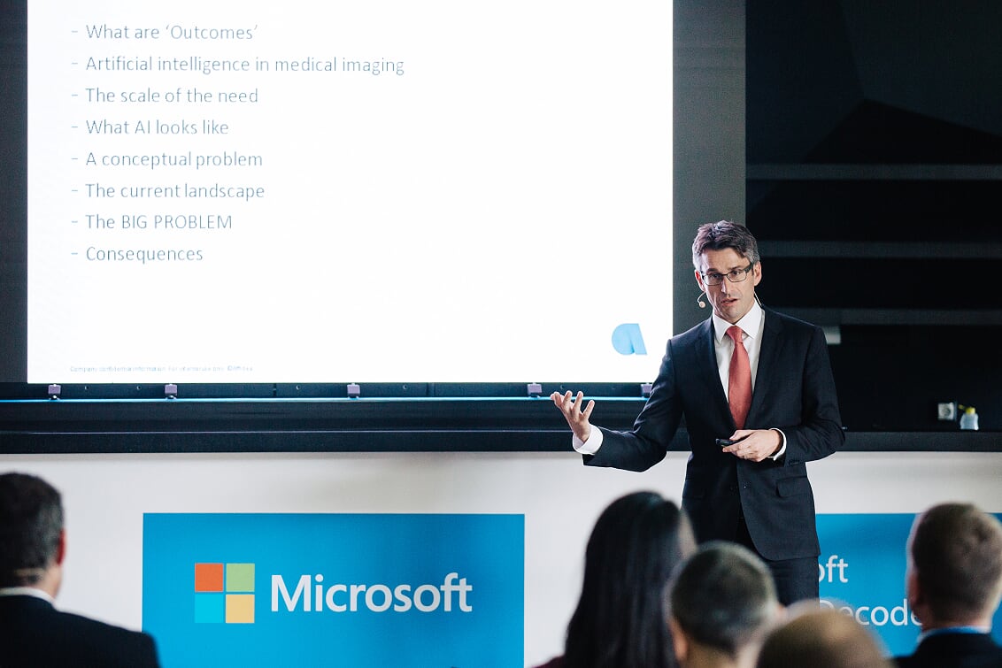
Just about everyone knows that it is not so easy for doctors to evaluate the images produced by medical imaging devices (ultrasound, x-ray, CT, MRI), but probably few people are aware of the real difficulties they face. A CT scan produces “only” a black and white image, but in these images there can be as many as 4,000 different shades of grey! Rowland Illing is Chief Medical Officer at Affidea, a company involved in diagnostics imaging analysis. At Microsoft’s Future Decoded event, he gave us an idea about the magnitude of the task: “The monitors used for assessing the images are able to display 256 of these shades, and the human eye is able to differentiate between 20 or 30.” (Dr Illing is a real hotshot in his specialist field. He teaches at several universities, including Semmelweis University, while also working as a consultant to the European Commission, Microsoft and IBM in the field of the medical use of AI.)
First the images are stored in one location, in the Microsoft Azure service. This enables Azure to analyse them with its cognitive service. Image evaluation is one of the specific fields where artificial intelligence may have a huge role. AI is able to detect all shades of grey, identifying each one precisely, and it does not get tired or bored. And there is a huge demand for it. According to Illing, medical scanning images are made of 450 million people every year globally, among which 90 million are misdiagnosed, while the problem remains undetected in 110 million cases. All this represents huge potential for AI. In the United Kingdom it is now a rule that all images must be examined by two doctors (and research says that the second doctor reaches a different conclusion in 30 percent of the cases). An obvious field of use would be for artificial intelligence to replace this second pair of eyes. Also there is no need to be afraid of radiologists losing their jobs: their number is not increasing in most places, but the demand for image evaluation is rising by 10 percent every year.
In the meantime AI is becoming increasingly precise. Dr Illing mentioned a real example: out of every one thousand tested women, five have breast cancer on average, but after the first test, every tenth is called back for a check-up. In other words, 95 women are made unnecessarily anxious and worried with unnecessary biopsies being performed on them. An international competition was announced for the development of an algorithm than can be used for reducing the number of false positive results. The winner reduced the number of false positives by five percent.
At the same time, the use of artificial intelligence in the evaluation of the images raises an interesting problem. If the doctor sees something on the image that the AI also views as a problem, the doctor is pleased. But what happens if the AI identifies something, but the doctor thinks there is nothing there? In this case whom should we believe? The machine, or the doctor’s own eyes and experience?
All things considered, Dr Illing is quite sure that artificial intelligence has a big future in medical imaging. Nor is he afraid of AI taking the place of the doctor. Because the future won’t be about people against AI, but people against people with AI—and it is obvious who will be on the winning side.
For Hungarian version please click the link below:
https://news.microsoft.com/hu-hu/2018/11/28/mesterseges-intelligencia-a-diagnosztikaban/

