Culture of Innovation
AI and X-rays: Identifying the many faces of COVID-19
Lunit's technology is helping doctors in many countries identify, triage and monitor patients.
 Steven Borowiec
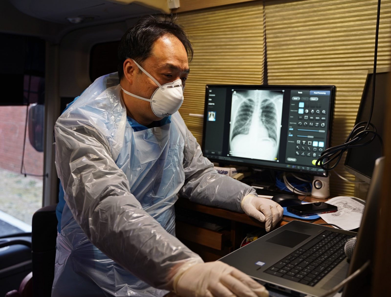
A digital diagnostic tool that uses artificial intelligence and cloud computing to accurately read vast numbers of chest X-rays – faster than a radiologist can – is helping doctors identify, triage and monitor COVID-19 patients.
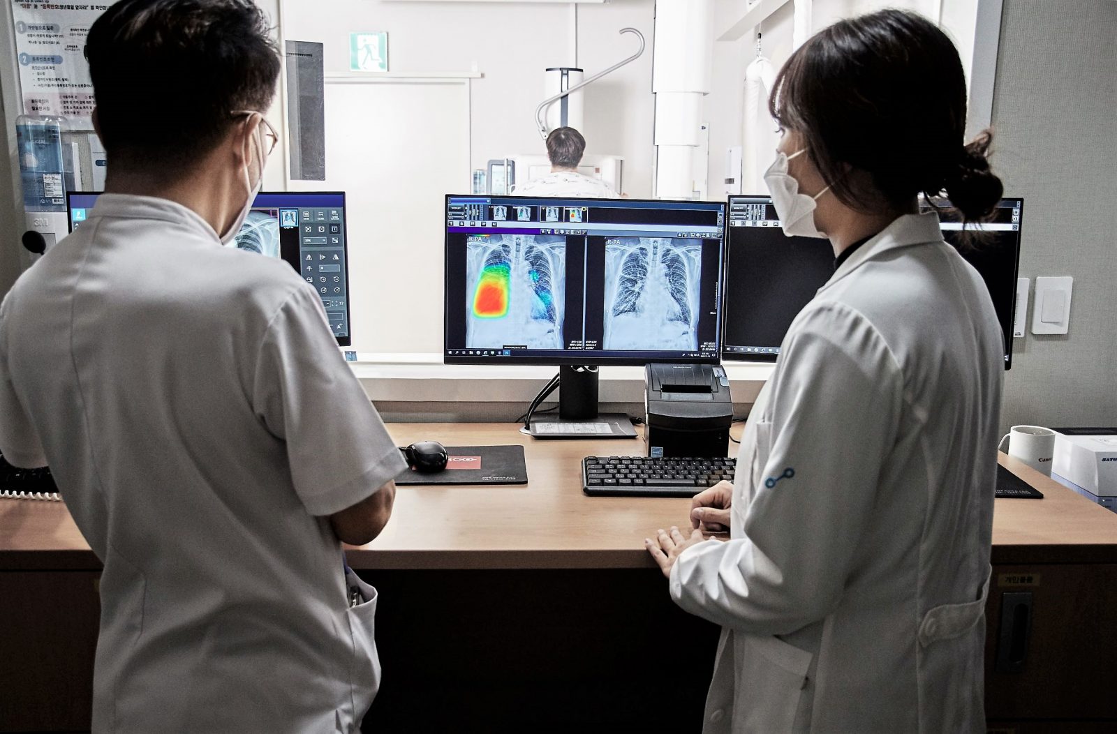
South Korean radiologist, Dr. Kyu-mok Lee, says the rapid analysis capabilities of Lunit INSIGHT CXR allow physicians to quickly identify what he describes as “the many faces of COVID-19” so they can expedite patient care and stem community transmission.
Identifying and isolating suspected patients quickly has become critical to controlling the spread of the coronavirus even as vaccination programs move ahead. But doing that can be a big challenge when testing processes, staffing and other resources are stretched by overflowing numbers of patients.
This is why hospitals, screening centers and clinics in multiple countries are increasingly turning to fast and accurate AI-aided chest X-ray exams for COVID-19 case triaging.
Lunit’s algorithms have been trained to read X-rays and originally could detect signs of 10 major chest diseases, including cancers, with an accuracy rate of 97% to 99%.
When the pandemic struck, its developers quickly adapted them to also scan for signs of COVID-19 – including pneumonia, which is often present in infected patients.
By innovating with agility, South Korean software company Lunit has created a valuable and fast support tool for medical professionals trying to manage pandemic cases.
Lee and his radiology team joined the COVID-19 frontline when the first outbreak hit Seoul about a year ago.
They initially worked out of a makeshift community treatment center at a major city hospital that had a cascading overflow of cases. Thousands of people were being swabbed and tested every day at the height of the outbreak.
Then as now, doctors needed accurate and fast ways to determine which patients had contracted the virus, and at what levels, so they could be appropriately isolated and treated.
Suspected COVID-19 patients can present with widely different symptoms at different levels of intensity. Some have tell-tale signs such as coughing, fever, fatigue and aches and pains. Some have mild cases and others severe, while a number appear asymptomatic.
Asymptomatic patients show no immediate signs of infection. But they can still shed the virus and unknowingly spread it to others, posing a potential threat to the wider community.
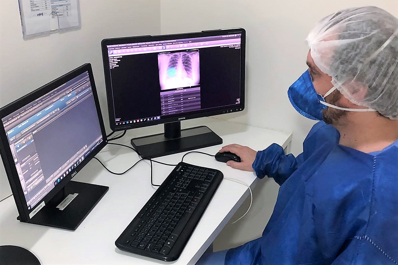
Along with South Korea, the technology has been adopted in Thailand, Indonesia, Mexico, Italy and France. In hard-hit Brazil, a major health group is using it to screen chest X-rays of patients with mild COVID-19 symptoms. Medical workers can triage cases faster and more effectively in hospitals with high patient numbers but few radiologists.
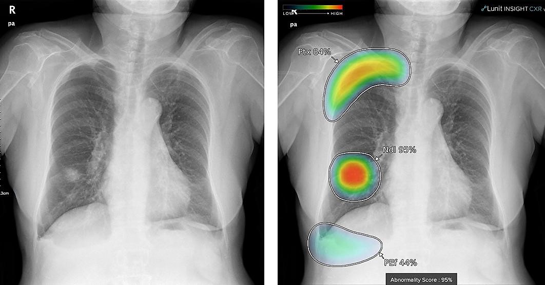
X-rays only appear in black and white, which means that there are cases where lesions aren’t noticeable to the human eye. Lunit’s solution has the advantage of displaying lesions in vivid color.
– Dr. Kyu-mok Lee, radiologist
Lunit’s technology not only helps doctors who are experts in reading X-ray images, but also those without extensive radiology skills. This has proven valuable during the pandemic.
“Physicians from various fields such as orthopedics, psychiatry, and family medicine, have been taking part in (COVID-19) treatment. They are not trained radiologists, so they are limited in their ability to read X-ray test results,” Lee said.
“X-rays only appear in black and white, which means that there are cases where lesions aren’t noticeable to the human eye. Lunit’s solution has the advantage of displaying lesions in vivid color, which makes them more noticeable.”
It can help medical staff make informed decisions. But a patient’s fate is not put solely in the hands of AI. Cases that have been flagged by the technology are followed up and the results are double-checked by doctors.
Lee says the processing scale and accuracy of Lunit’s technology can help free up busy radiologists.
“An X-ray is a compressed two-dimensional rendering of three-dimensional human structures. Inevitably, organs and structures overlap in the images, which can make it easy for the human eye to miss lesions,” he said.
“The reality for radiologists, especially in Korea, is that it’s impossible to invest a lot of time in reading each X-ray as they would have to read hundreds or thousands every day.”
Using the cloud computing power of Microsoft Azure, Lunit’s technology generates location information of detected lesions in the form of heatmaps. It also makes abnormality scores that reflect the probability that the detected lesion is abnormal and needs further investigation by a radiologist.
Even seasoned radiologists can sometimes miss vital details when under pressure. Lee recalls a recent case where Lunit’s algorithms found a COVID-19-related lesion in a patient’s lung that had gone undetected by doctors.
If this had remained unnoticed and untreated, the patient would have developed a far more serious case that would have meant hospitalization. “Detecting the lesion early made a real difference,” Lee says.
Chest radiography is still a supplementary tool to the more definitive but far slower polymerase chain reaction test for COVID, Lee says. But Lunit’s solution has made an impact in efforts to stem the outbreak.
Lunit’s software is continuously being improved, having analyzed more than 6.5 million chest X-ray images from more than 80 countries around the world.
Data collected from diagnoses is also being used for research and training. Patient privacy is protected as all information is stripped of personal identification.
![]() Lunit’s technology has received recognition through studies published in major peer-reviewed journals such as Radiology, Lancet Digital Health, JAMA Network Open, Clinical Infectious Diseases and others.
Lunit’s technology has received recognition through studies published in major peer-reviewed journals such as Radiology, Lancet Digital Health, JAMA Network Open, Clinical Infectious Diseases and others.
By enabling more accurate, efficient, and timely diagnosis of chest diseases, Lunit INSIGHT CXR can help reduce the workload of medical professionals. In this way, they can bring more value to the patients.
– Brandon Suh, CEO, Lunit
Lunit was founded as a company in 2013 to make AI- and data-driven medicine a new standard of care.
“We are especially focused on conquering cancer, one of the leading causes of death worldwide,” says CEO Brandon Suh. “We develop AI solutions for precision diagnostics and therapeutics, to find the right diagnosis at the right cost, and the right treatment for the right patients.
“By enabling more accurate, efficient, and timely diagnosis of chest diseases, Lunit INSIGHT CXR can help reduce the workload of medical professionals. In this way, they can bring more value to the patients in not only difficult circumstances, such as the current pandemic crisis, but in routine clinical settings as well.”
Meanwhile, the company has made major advances in the AI-driven diagnosis of other diseases. For instance, another solution known as Lunit INSIGHT MMG, is as accurate as an average radiologist in identifying breast cancer in mammograms, recent studies show.
By reaching this milestone, its developers think it could potentially have a role as an independent image reader to reduce radiologist workload and possibly increase cancer detection in the future.
TOP IMAGE courtesy of Lunit